
Plasmodium vivax: Thin Blood Smear
Original bench-card visuals for recognizing Plasmodium vivax on a Giemsa-stained thin smear: enlarged red cells, Schuffner dots, ameboid trophozoites, schizonts with 12-24 merozoites, and round gametocytes.
Plasmodium vivax: enlarged RBC, Schuffner dots, ameboid trophozoite
P. vivax prefers young red cells, so infected cells are enlarged, often up to 1.5-2 times normal. Schuffner dots (fine pink stippling) appear as the parasite grows. All stages circulate: ring, ameboid trophozoite, schizont with 12...
The ring is about one third of the red cell diameter with a single large chromatin dot. Early rings cannot be separated reliably from other species, so look for olde...
The growing trophozoite has irregular, spread-out cytoplasm, described as ameboid. The red cell is clearly enlarged and shows fine pink Schuffner dots. Fine golden-b...
A mature schizont holds 12-24 merozoites and nearly fills the enlarged cell, with clumped pigment. This count is higher than in P. malariae or P. ovale.
Round to oval and filling most of the enlarged cell, with scattered pigment. Never banana shaped; a crescent gametocyte points to P. falciparum.
What this looks like down the scope
The board above is drawn to isolate the pattern. These are clinical images of the same stages, published as you see them, so you can find the pattern with everything else still in the field.
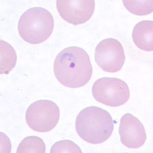
Thin film, Giemsa. Large rings in cells already bigger and paler than their neighbours. The enlarged host cell is the first vivax clue, before you judge the parasite at all.
Source: CDC / DPDx | Public domain (U.S. federal government work) | Original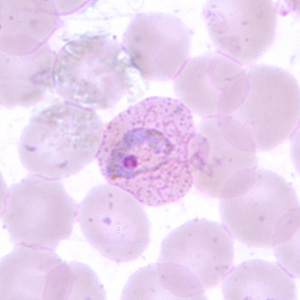
Thin film. The trophozoite sprawls ameboid-fashion inside an enlarged cell, with fine Schuffner's stippling across the cytoplasm. Vivax invades young cells, which is why the host cell swells.
Source: CDC / DPDx | Public domain (U.S. federal government work) | Original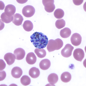
Thin film. A mature schizont crowded with merozoites, typically 12 to 24 of them. That is more than any other species you will meet on a routine film.
Source: CDC / DPDx | Public domain (U.S. federal government work) | OriginalShown as published and free of charge from the source above, which does not endorse Learn Microbes. Full image credits
Thin smear anchors
| Visual clue | Meaning | Student shorthand |
|---|---|---|
| Enlarged infected RBC | P. vivax or P. ovale | P. falciparum and P. malariae do not enlarge the cell |
| Schuffner dots | P. vivax or P. ovale | Seen best when the stain buffer is near pH 7.0-7.2 |
| Ameboid trophozoite | P. vivax | P. ovale and P. malariae trophozoites are compact |
| Schizont with 12-24 merozoites | P. vivax | P. ovale and P. malariae usually have 6-12 |
| All stages present in peripheral blood | Not typical P. falciparum | P. falciparum usually shows only rings and gametocytes |
Plasmodium vivax study readout
| Pattern | What to call it | How to think about it |
|---|---|---|
| Enlarged RBC, Schuffner dots, ameboid trophozoite | Plasmodium vivax | Report species and percent parasitemia per SOP |
| Enlarged oval RBC with ragged edge, compact trophozoite | Consider Plasmodium ovale | Compare with the P. ovale bench card |
| Rings only, normal-size RBC | Species not established | Search more fields for older stages; rule out P. falciparum first |
- Enlarged red cell plus Schuffner dots narrows the choice to P. vivax or P. ovale.
- Ameboid trophozoites and 12-24 merozoites favor P. vivax.
- A positive malaria smear is a critical result. Species and parasitemia follow your lab policy.
Big cell, pink dots, ameboid parasite, up to 24 merozoites = P. vivax.
New bench cards and study tools are added regularly. Join the Lab List to hear about new cards and have a say in what gets built next.