
Nonpathogenic Amoeba Cysts: Entamoeba coli, Endolimax nana, Iodamoeba butschlii
Original comparison visuals for the three commensal amoeba cysts students confuse with Entamoeba histolytica: Entamoeba coli with up to eight nuclei, tiny Endolimax nana with four blot-like karyosomes, and Iodamoeba butschlii with its large glycogen vacuole.
Commensal amoeba cysts: count the nuclei, read the karyosome, check for a vacuole
Entamoeba coli: about 10-35 um, up to 8 nuclei when mature, eccentric karyosome, coarse uneven peripheral chromatin. Endolimax nana: about 5-10 um, oval, 4 nuclei with large blot-like karyosomes and no peripheral chromatin. Iodamo...
About 10-35 um, round. A mature cyst has 8 nuclei, more than any Entamoeba histolytica cyst. Each nucleus has an eccentric karyosome and coarse, uneven peripheral ch...
About 5-10 um, usually oval. Four nuclei, each showing a large blot-like karyosome with no peripheral chromatin, so the nuclei look like four dark dots. One of the s...
About 5-20 um, often irregular in shape. One nucleus with a large karyosome. The large, sharply outlined glycogen vacuole stains deep brown with iodine and appears a...
What this looks like down the scope
The board above is drawn to isolate the pattern. These are clinical images of the same stages, published as you see them, so you can find the pattern with everything else still in the field.
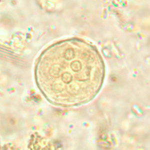
Six nuclei in this plane alone. Rack the focus and more appear - up to eight in a mature cyst, which no pathogen in this group reaches.
Source: CDC / DPDx | Public domain (U.S. federal government work) | Original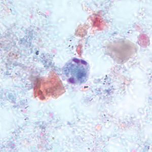
E. hartmanni: built like E. histolytica but under 10 um. Size alone separates them, so this is a call that needs a measured eyepiece, not an impression.
Source: CDC / DPDx | Public domain (U.S. federal government work) | Original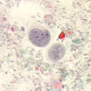
Endolimax nana cysts, small and oval. The karyosomes are large, irregular blots that fill much of each nucleus, and there is no peripheral chromatin at all.
Source: CDC / DPDx | Public domain (U.S. federal government work) | Original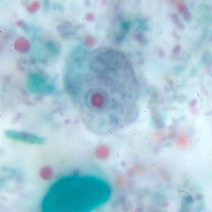
The trophozoite, with that same blot-like karyosome. A nucleus with no rim of chromatin is the quickest way to rule Entamoeba out.
Source: CDC / DPDx | Public domain (U.S. federal government work) | Original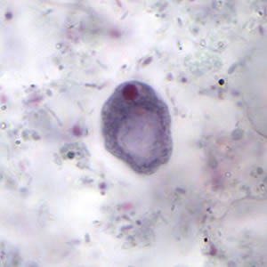
Iodamoeba butschlii. The dark mass taking up most of the cyst is a glycogen vacuole - large, single, and present in no other cyst here.
Source: CDC / DPDx | Public domain (U.S. federal government work) | Original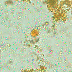
The same species in iodine, which is where the name comes from: iodine stains that glycogen mass a deep brown.
Source: CDC / DPDx | Public domain (U.S. federal government work) | OriginalShown as published and free of charge from the source above, which does not endorse Learn Microbes. Full image credits
Cyst differential anchors
| Visual clue | Meaning | Student shorthand |
|---|---|---|
| More than 4 nuclei in an Entamoeba cyst | Entamoeba coli | E. histolytica/dispar cysts stop at 4 |
| Eccentric karyosome, coarse uneven chromatin | Entamoeba coli nucleus | E. histolytica: central karyosome, fine even chromatin |
| Splinter-shaped chromatoid bodies | Entamoeba coli | E. histolytica chromatoid bodies have rounded ends |
| Tiny oval cyst with 4 dark dots | Endolimax nana | No peripheral chromatin on the nuclear membrane |
| Single large iodine-brown vacuole | Iodamoeba butschlii | Vacuole is clear and unstained on trichrome |
Commensal amoeba cyst readout
| Pattern | What to call it | How to think about it |
|---|---|---|
| Cyst with 8 nuclei, eccentric karyosomes | Entamoeba coli | Nonpathogen; report per SOP and keep looking for pathogens |
| Small oval cyst, 4 blot-like karyosomes | Endolimax nana | Nonpathogen; smallest common amoeba cyst |
| Cyst with one nucleus and a large glycogen vacuole | Iodamoeba butschlii | Nonpathogen; iodine makes the vacuole obvious |
- Eight nuclei rule out Entamoeba histolytica and point to Entamoeba coli.
- Endolimax nana and Iodamoeba butschlii have large karyosomes with no peripheral chromatin.
- Finding a commensal does not end the exam. Mixed infections with pathogens are common.
8 nuclei = E. coli. 4 dots in a tiny oval = E. nana. Big iodine-brown vacuole = Iodamoeba.
New bench cards and study tools are added regularly. Join the Lab List to hear about new cards and have a say in what gets built next.