
Microsporidia: Spores on Modified Trichrome Stain
Original bench-card visuals for recognizing microsporidia spores in stool on a modified trichrome stain: tiny pink-red oval spores, some with a belt-like stripe, compared with larger budding yeast.
Microsporidia spores: tiny, pink-red, some with a belt-like stripe
Microsporidia spores are only about 1-4 um, close to the size of bacteria, so they are read under oil immersion. On a modified trichrome stain (high chromotrope 2R), spores stain pink-red against a green or blue background. Some s...
Oval spores about 1-4 um with a pink-red wall. Some show a horizontal or diagonal stripe, which is the coiled polar tubule seen through the wall. Enterocytozoon bien...
Yeast cells also take up the red stain but are larger, commonly 3-6 um or more, and show budding. They have no belt-like stripe. Bacteria may stain as well but are u...
What this looks like down the scope
The board above is drawn to isolate the pattern. These are clinical images of the same stages, published as you see them, so you can find the pattern with everything else still in the field.
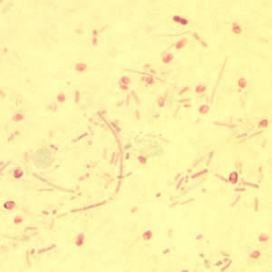
Chromotrope 2R. The spores are tiny pink-red ovals, 1-3 um - near the limit of what light microscopy can resolve, which is why this stain exists.
Source: CDC / DPDx | Public domain (U.S. federal government work) | Original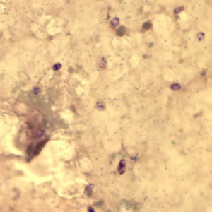
Gram chromotrope on the same kind of specimen, which darkens the spores and makes them easier to pick out of debris.
Source: CDC / DPDx | Public domain (U.S. federal government work) | Original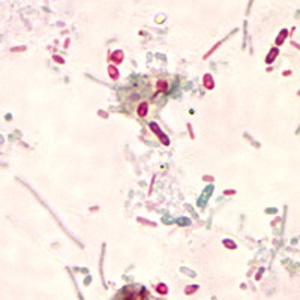
Spores among debris, which is the realistic view. Look for the uniform size and the crisp oval outline; bacteria and yeast in the same field are neither.
Source: CDC / DPDx | Public domain (U.S. federal government work) | OriginalShown as published and free of charge from the source above, which does not endorse Learn Microbes. Full image credits
Modified trichrome anchors
| Visual clue | Meaning | Student shorthand |
|---|---|---|
| Tiny oval pink-red bodies, 1-4 um | Possible microsporidia spores | Confirm with oil immersion and controls |
| Belt-like stripe across the spore | Strong clue for microsporidia | Represents the polar tubule |
| Budding cells, larger size | Yeast | Common in stool; do not report as spores |
| Chronic watery diarrhea, immunocompromised patient | Clinical setting that raises suspicion | The stain must be requested or reflexed; the routine O&P misses it |
| Bright spores with an optical brightener | Chitin wall stained | Sensitive but not specific; yeast fluoresces too |
Microsporidia study readout
| Pattern | What to call it | How to think about it |
|---|---|---|
| Pink-red oval spores, 1-4 um, some with a stripe; control acceptable | Microsporidia spores detected | Report per SOP; species requires reference methods |
| Red-staining budding cells of larger size | Yeast, not microsporidia | Do not report as spores |
| Brightener-positive small ovals only | Screen positive | Confirm with modified trichrome before reporting |
- Spores are bacteria-sized, so oil immersion and a special stain are required.
- Pink-red oval spores with a belt-like stripe on modified trichrome are the classic finding.
- Size and budding separate yeast from microsporidia.
Tiny pink-red ovals with a belt stripe on modified trichrome = microsporidia. Routine O&P will miss them.
New bench cards and study tools are added regularly. Join the Lab List to hear about new cards and have a say in what gets built next.