
Candida on Cornmeal Agar: Species Morphology Comparison
Original comparison visuals for reading yeast morphology on cornmeal agar with Tween 80: Candida albicans chlamydospores, Candida tropicalis blastoconidia along long pseudohyphae, Candida parapsilosis fine curved pseudohyphae, and Candida glabrata with yeast cells only.
Cornmeal agar (Dalmau) morphology: four Candida patterns
The yeast is streaked and cut into cornmeal agar with Tween 80, covered with a coverslip, incubated at room temperature for 48-72 hours, and read through the coverslip on low and high dry power. Look for three things in order: are...
Pseudohyphae and true hyphae with dense round clusters of blastoconidia at the septa, plus large, round, thick-walled chlamydospores at the tips. Chlamydospores are...
Abundant long pseudohyphae with blastoconidia placed singly or in small groups anywhere along them, not in tight clusters. No chlamydospores in the usual picture.
Thin, short, curved pseudohyphae that branch off the streak line, often described as sagebrush or spider-like. Occasional large, wide pseudohyphal cells called giant...
Small oval budding yeast cells, about 2-4 um, with no pseudohyphae and no chlamydospores. Now also published as Nakaseomyces glabratus. Cryptococcus also lacks pseud...
What this looks like down the scope
The board above is drawn to isolate the pattern. These are clinical images of the same stages, published as you see them, so you can find the pattern with everything else still in the field.
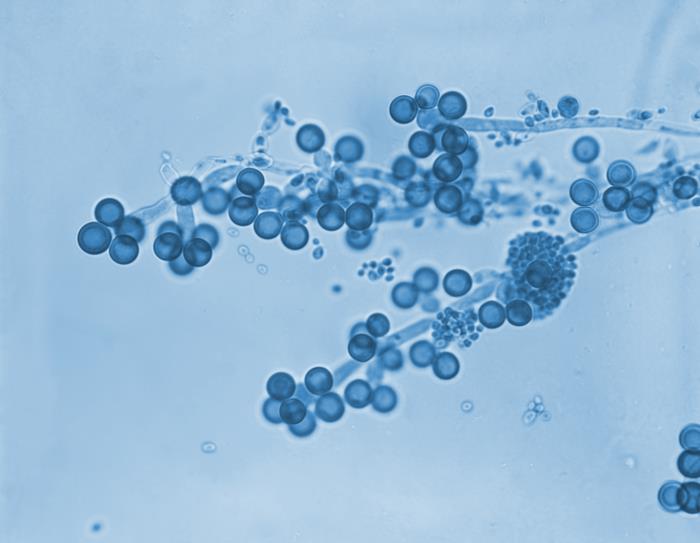
Cornmeal agar at 475x. The large round thick-walled bodies at the ends of the pseudohyphae are chlamydospores, and clusters of small blastoconidia sit at the joints. Only C. albicans and C. dubliniensis make these.
Source: CDC / PHIL - Dr. Lucille K. Georg | Public domain (U.S. federal government work) | Original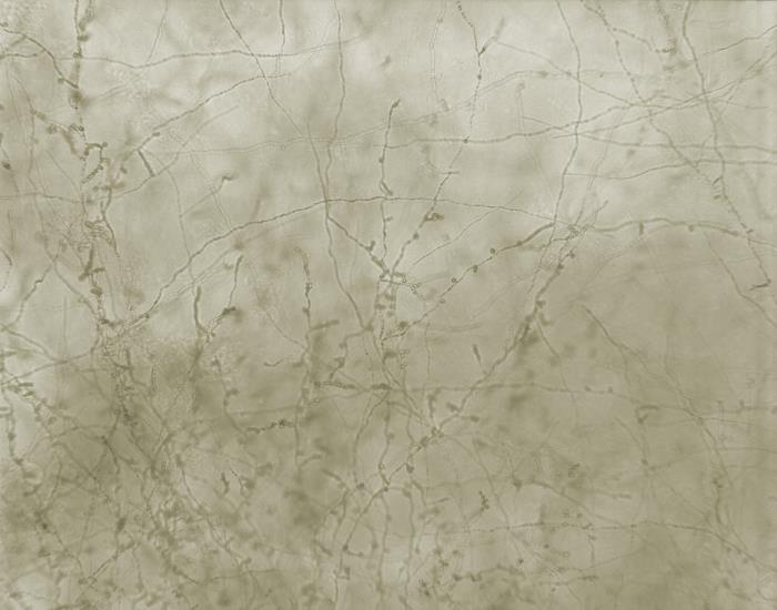
Long branching pseudohyphae running across the field with blastoconidia scattered singly or in small groups rather than in tight clusters. No chlamydospores anywhere.
Source: CDC / PHIL - Dr. Hilliard F. Hardin | Public domain (U.S. federal government work) | Original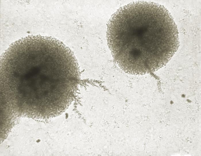
Two microcolonies with fine pseudohyphae spraying out from the edge, curved and short. Compare the delicacy against the C. tropicalis frame: same structure, very different build.
Source: CDC / PHIL - Dr. Hilliard F. Hardin | Public domain (U.S. federal government work) | OriginalShown as published and free of charge from the source above, which does not endorse Learn Microbes. Full image credits
Cornmeal agar anchors
| Visual clue | Meaning | Student shorthand |
|---|---|---|
| Terminal thick-walled chlamydospores | C. albicans or C. dubliniensis | Pair with a germ tube test |
| Blastoconidia in tight clusters at septa | C. albicans pattern | Looks like grape bunches at regular intervals |
| Blastoconidia scattered singly along long pseudohyphae | C. tropicalis pattern | No chlamydospores |
| Fine curved pseudohyphae, giant cells | C. parapsilosis pattern | Sagebrush or spider-like growth |
| Small yeast only, no pseudohyphae | C. glabrata pattern | Rule out Cryptococcus with urease |
Cornmeal morphology study readout
| Pattern | What to call it | How to think about it |
|---|---|---|
| Chlamydospores present, germ tube positive | Candida albicans (or C. dubliniensis) | Most labs report C. albicans by rapid tests and confirm when needed |
| Pseudohyphae present, no chlamydospores | Non-albicans Candida | Read blastoconidia arrangement and continue identification |
| Yeast cells only | C. glabrata, Cryptococcus, or another non-filamenting yeast | Urease and further testing separate them |
- Read cornmeal agar in order: pseudohyphae, blastoconidia arrangement, chlamydospores.
- Chlamydospores point to C. albicans; yeast cells alone point to C. glabrata.
- A true germ tube has no constriction at its origin.
Chlamydospores = C. albicans. No pseudohyphae at all = think C. glabrata.
New bench cards and study tools are added regularly. Join the Lab List to hear about new cards and have a say in what gets built next.