
Yeast Forms in Tissue: Size and Budding Comparison
Original comparison visuals for the systemic fungi as they appear in tissue and body fluids: small intracellular Histoplasma, encapsulated Cryptococcus, broad-based budding Blastomyces, and the large Coccidioides spherule.
Tissue forms compared: size first, then how the cell buds
In a direct smear or tissue section, two questions separate most systemic fungi: how big is the cell, and how does it bud. Histoplasma capsulatum: 2-4 um, inside macrophages, narrow-based bud. Cryptococcus: 4-10 um with wide size...
Small oval yeast cells packed inside a macrophage. A clear halo around each cell is a staining artifact and not a true capsule. Budding is on a narrow base.
Round yeast cells that vary a lot in size within one field. Narrow-based budding. The polysaccharide capsule shows as a clear zone on India ink and stains with mucic...
Large round yeast with a thick, double-contoured wall and a single bud attached by a wide base. The bud stays attached until it is nearly the size of the mother cell...
A thick-walled spherule, commonly 30-60 um and up to about 100 um, filled with endospores 2-5 um across. It does not bud. A ruptured spherule releases endospores tha...
What this looks like down the scope
The board above is drawn to isolate the pattern. These are clinical images of the same stages, published as you see them, so you can find the pattern with everything else still in the field.
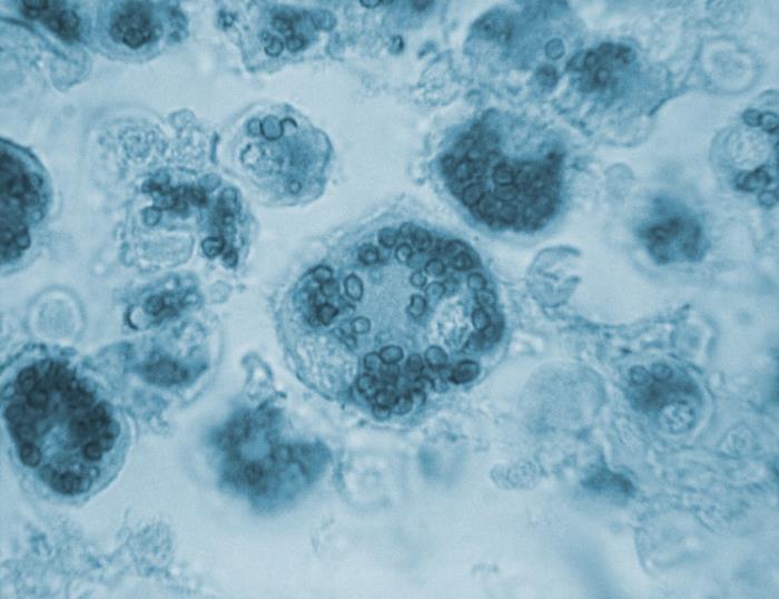
Smallest, at 2-4 um, and almost always inside a host cell. If you are looking at free yeasts in the open field, you are probably not looking at Histoplasma.
Source: CDC / PHIL - Dr. Lucille K. Georg | Public domain (U.S. federal government work) | Original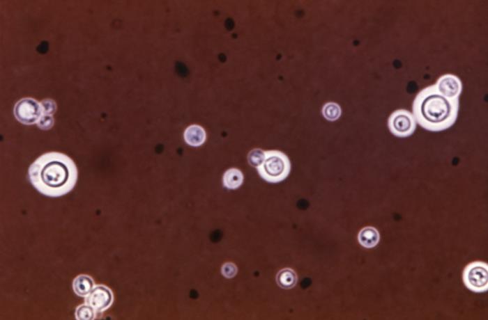
Next up at 4-10 um, and the only one on this card with a capsule. Cell size varies a lot within one field, which none of the others do.
Source: CDC / PHIL - Sherry A. Brinkman | Public domain (U.S. federal government work) | Original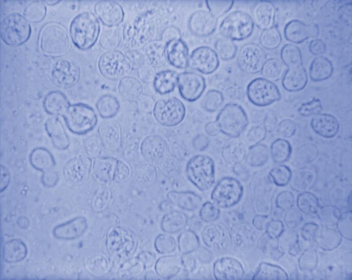
Larger again at 8-15 um, with a wall thick enough to read as a double line and buds joined across a broad neck. Size plus wall thickness plus the broad bud, in that order.
Source: CDC / PHIL - Dr. Lucille K. Georg | Public domain (U.S. federal government work) | Original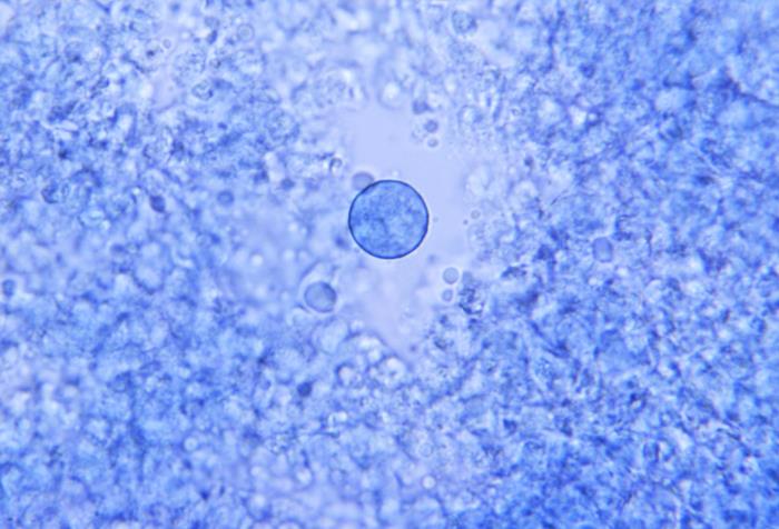
Not a yeast at all. A spherule reaches 20-60 um or more and is filled with endospores, and it never buds - so if you cannot find a bud anywhere, stop and measure.
Source: CDC / PHIL - Dr. Lucille K. Georg | Public domain (U.S. federal government work) | OriginalShown as published and free of charge from the source above, which does not endorse Learn Microbes. Full image credits
Tissue form anchors
| Visual clue | Meaning | Student shorthand |
|---|---|---|
| 2-4 um yeasts inside macrophages | Histoplasma capsulatum | Bone marrow, blood buffy coat, lung, or lymph node |
| Round yeasts of mixed sizes with a capsule | Cryptococcus | CSF India ink; cryptococcal antigen is more sensitive |
| Thick-walled yeast with one broad-based bud | Blastomyces | Figure-eight appearance |
| Large spherule with endospores | Coccidioides | No budding at any stage |
| Large cell with many narrow-based buds around it | Paracoccidioides | Mariner's wheel; see its bench card |
Tissue yeast study readout
| Pattern | What to call it | How to think about it |
|---|---|---|
| Small intracellular yeasts, narrow-based budding | Consistent with Histoplasma capsulatum | Correlate with antigen testing and culture |
| Encapsulated yeasts, variable size | Consistent with Cryptococcus | Correlate with cryptococcal antigen |
| Broad-based budding yeasts or spherules with endospores | Consistent with Blastomyces or Coccidioides | Notify per policy; cultures need containment |
- Size and budding pattern separate the systemic fungi in direct specimens.
- Narrow base: Histoplasma and Cryptococcus. Broad base: Blastomyces. No budding: Coccidioides.
- Report direct findings as consistent with an organism and confirm by another method.
Small and intracellular = Histoplasma. Capsule = Cryptococcus. Broad base = Blastomyces. Spherule = Coccidioides.
New bench cards and study tools are added regularly. Join the Lab List to hear about new cards and have a say in what gets built next.