
Pneumocystis jirovecii: Cysts on GMS and Trophic Forms on Giemsa
Original bench-card visuals for recognizing Pneumocystis jirovecii in respiratory specimens: cup-shaped and crescent cysts on silver stain, and clusters of trophic forms in foamy material on Giemsa.
Pneumocystis jirovecii: silver-stained cysts and Giemsa-stained clusters
Pneumocystis jirovecii is a fungus that cannot be grown in routine culture, so diagnosis depends on stains, immunofluorescence, or molecular testing of respiratory specimens such as bronchoalveolar lavage or induced sputum. On Gom...
Cysts are about 4-7 um with a dark brown-black wall on a green background. Collapsed cysts look like cups, crescents, or crushed ping-pong balls. Many show a dark co...
Giemsa or a rapid Giemsa-type stain shows clusters of trophic forms about 1-4 um, with small purple nuclei in pale blue cytoplasm, set in foamy material. Cyst walls...
What this looks like down the scope
The board above is drawn to isolate the pattern. These are clinical images of the same stages, published as you see them, so you can find the pattern with everything else still in the field.
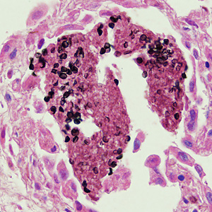
Methenamine silver turns the cyst wall black and leaves everything else pink. Look for round, cup-shaped and crescent outlines, some collapsed - and never a bud, because this organism does not bud.
Source: CDC / DPDx | Public domain (U.S. federal government work) | Original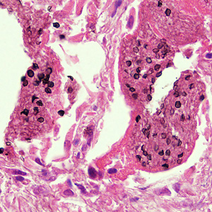
A wider field. The cysts sit inside the frothy alveolar exudate rather than in the tissue itself, which is where to look first on a low-power scan.
Source: CDC / DPDx | Public domain (U.S. federal government work) | Original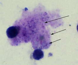
The arrows are the source's own. Giemsa stains the trophozoite nuclei as small dots inside a foamy mass and leaves the cyst wall unstained, so there is nothing with an outline to find - which is exactly why CDC marked them.
Source: CDC / DPDx | Public domain (U.S. federal government work) | OriginalShown as published and free of charge from the source above, which does not endorse Learn Microbes. Full image credits
Stain anchors
| Visual clue | Meaning | Student shorthand |
|---|---|---|
| Cup or crescent black cysts on GMS | Pneumocystis jirovecii cysts | Collapsed cyst shapes are the classic picture |
| No budding | Pneumocystis, not a yeast | Histoplasma and Candida bud |
| Foamy clusters on Giemsa | Trophic forms and intracystic bodies | The cyst wall itself is unstained |
| Apple-green cysts with fluorescent antibody | Direct fluorescent antibody positive | More sensitive than GMS or Giemsa |
| Positive PCR, negative stain | Detected at low level | Infection versus colonization needs clinical judgment |
Pneumocystis study readout
| Pattern | What to call it | How to think about it |
|---|---|---|
| Clustered non-budding cysts, cup and crescent forms on GMS | Pneumocystis jirovecii detected | Report per SOP; this is a significant result in an immunocompromised patient |
| Budding oval cells of similar size | Yeast, not Pneumocystis | Consider Histoplasma or Candida; correlate with culture and antigen tests |
| No cysts in an adequate lavage specimen | No Pneumocystis seen | A negative stain does not exclude infection; PCR may be requested |
- Pneumocystis is a fungus that is not recovered in routine culture.
- GMS stains the cyst wall; Giemsa stains the trophic forms and intracystic bodies.
- No budding plus cup and crescent shapes separates it from small yeasts.
Cup-shaped, non-budding black cysts on GMS in a lavage = Pneumocystis jirovecii.
New bench cards and study tools are added regularly. Join the Lab List to hear about new cards and have a say in what gets built next.