
Plasmodium malariae: Band Form and Rosette Schizont
Original bench-card visuals for recognizing Plasmodium malariae on a thin smear: normal-size or small red cells, band-form trophozoites, rosette schizonts with 6-12 merozoites, and coarse dark pigment.
Plasmodium malariae: band trophozoite and rosette schizont in a normal-size RBC
P. malariae prefers older red cells, so infected cells are normal in size or slightly small, with no Schuffner dots. The trophozoite is compact and may stretch across the cell as a band. The mature schizont has 6-12 merozoites, of...
The compact trophozoite stretches from one side of the red cell to the other as a band, with coarse dark pigment along it. The red cell is not enlarged and has no st...
The mature schizont has 6-12 merozoites, commonly about 8, arranged in a ring around a central clump of pigment. This daisy or rosette pattern is the classic picture...
Round to oval, filling a red cell that stays normal in size. Abundant coarse dark pigment. It resembles a P. vivax gametocyte except that the host cell is not enlarg...
What this looks like down the scope
The board above is drawn to isolate the pattern. These are clinical images of the same stages, published as you see them, so you can find the pattern with everything else still in the field.
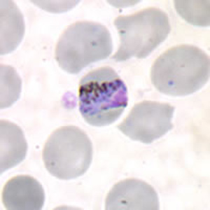
Thin film, Giemsa. The trophozoite stretched as a band straight across the cell, carrying coarse dark pigment. The host cell stays normal size or smaller, never enlarged.
Source: CDC / DPDx | Public domain (U.S. federal government work) | Original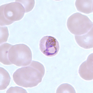
Thin film. A mature schizont with the merozoites set around a clump of central pigment, the rosette or basket arrangement. The count is low, usually 6 to 12.
Source: CDC / DPDx | Public domain (U.S. federal government work) | OriginalShown as published and free of charge from the source above, which does not endorse Learn Microbes. Full image credits
Thin smear anchors
| Visual clue | Meaning | Student shorthand |
|---|---|---|
| Normal or small infected RBC, no stippling | P. malariae or P. falciparum | Enlargement points to P. vivax or P. ovale |
| Band-form trophozoite | P. malariae | Also seen with P. knowlesi; check travel history |
| Rosette schizont with 6-12 merozoites | P. malariae | P. vivax has 12-24 |
| Coarse dark brown pigment | P. malariae | Heavier pigment than the other species |
| Low parasitemia, all stages present | Typical for P. malariae | Long 72-hour cycle, sometimes called quartan malaria |
Plasmodium malariae study readout
| Pattern | What to call it | How to think about it |
|---|---|---|
| Band forms, rosette schizonts, normal-size RBC | Plasmodium malariae | Report species and percent parasitemia per SOP |
| Band forms with high parasitemia, Southeast Asia travel | Consider Plasmodium knowlesi | Refer for molecular confirmation |
| Rings only in normal-size RBC | Species not established | Rule out P. falciparum before anything else |
- No cell enlargement and no Schuffner dots.
- Band-form trophozoites and rosette schizonts with 6-12 merozoites are the classic clues.
- A 72-hour cycle and low parasitemia fit P. malariae; high counts with band forms should make you think of P. knowlesi.
Normal-size cell + band form + daisy-head schizont = P. malariae.
New bench cards and study tools are added regularly. Join the Lab List to hear about new cards and have a say in what gets built next.