
Dermatophyte Hair Invasion: Ectothrix, Endothrix, and Favus
Original bench-card visuals for reading a KOH preparation of an infected hair: arthroconidia forming a sheath outside the shaft (ectothrix), arthroconidia packed inside the shaft (endothrix), and hyphae with air spaces (favus).
Hair invasion patterns: outside the shaft, inside the shaft, or hyphae with air spaces
Pluck hairs with the root attached, clear them in KOH, and look at where the fungal elements sit. Ectothrix: arthroconidia coat the outside of the shaft and the cuticle is damaged; typical of Microsporum canis and Microsporum audo...
Small arthroconidia form a mosaic sheath around the outside of the hair, and the cuticle is broken down. Microsporum canis and M. audouinii give this pattern and flu...
Arthroconidia fill the inside of the hair shaft in chains while the cuticle remains intact. The weakened hair snaps at the scalp surface, leaving black dots. Trichop...
Loose hyphae run along the inside of the shaft with air spaces or bubbles where hyphae have broken down. Arthroconidia are few. Caused by Trichophyton schoenleinii,...
What this looks like down the scope
The board above is drawn to isolate the pattern. These are clinical images of the same stages, published as you see them, so you can find the pattern with everything else still in the field.
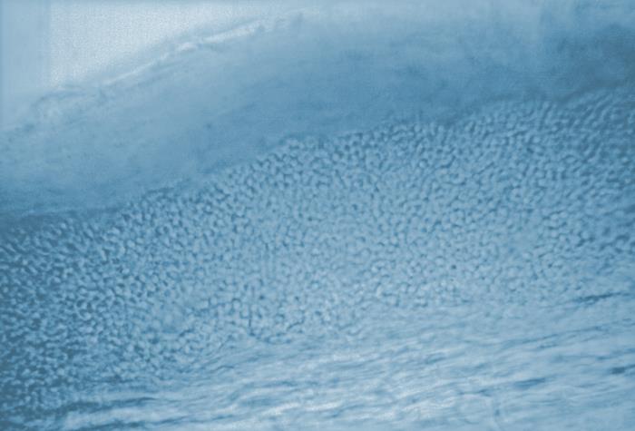
The spores form a sleeve on the outside of the shaft and the shaft itself stays clear. Ectothrix, and in Microsporum infections this is also where the Wood lamp fluorescence comes from.
Source: CDC / PHIL - Dr. Lucille K. Georg | Public domain (U.S. federal government work) | Original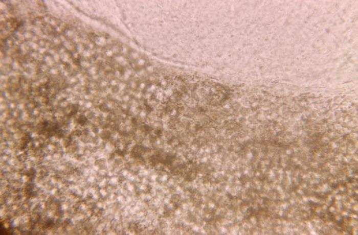
The same question answered the other way at 475x: the spores are inside the shaft, filling it, and the outer surface is bare. Endothrix. The hair breaks at the scalp because the shaft has been hollowed out.
Source: CDC / PHIL - Dr. Lucille K. Georg | Public domain (U.S. federal government work) | OriginalShown as published and free of charge from the source above, which does not endorse Learn Microbes. Full image credits
Hair invasion anchors
| Visual clue | Meaning | Student shorthand |
|---|---|---|
| Conidia outside the shaft, cuticle damaged | Ectothrix | Microsporum canis, M. audouinii, and others |
| Conidia inside the shaft, cuticle intact | Endothrix | Trichophyton tonsurans, T. violaceum |
| Hyphae and air spaces inside the shaft | Favus | Trichophyton schoenleinii |
| Yellow-green fluorescence under a Wood lamp | Microsporum canis or M. audouinii | A negative lamp does not rule out ringworm |
| Black dots at the scalp surface | Endothrix infection | Hairs break at the follicle opening |
Hair invasion study readout
| Pattern | What to call it | How to think about it |
|---|---|---|
| Ectothrix pattern, fluorescent hairs | Suggests Microsporum canis or M. audouinii | Culture confirms |
| Endothrix pattern, no fluorescence | Suggests Trichophyton tonsurans or T. violaceum | Culture confirms |
| Hyphae with air spaces | Favus pattern, Trichophyton schoenleinii | Culture confirms |
- Ectothrix means outside the shaft; endothrix means inside the shaft.
- Fluorescence under a Wood lamp points to certain Microsporum species only.
- The direct hair exam narrows the list and culture names the species.
Outside = ectothrix (Microsporum, fluoresces). Inside = endothrix (T. tonsurans, no fluorescence).
New bench cards and study tools are added regularly. Join the Lab List to hear about new cards and have a say in what gets built next.