
Free-Living Amoebae: Naegleria fowleri and Acanthamoeba
Original bench-card visuals for the free-living amoebae a lab must not miss: Naegleria fowleri trophozoites in CSF, and Acanthamoeba trophozoites and double-walled cysts in corneal or tissue specimens.
Naegleria fowleri and Acanthamoeba: trophozoites, flagellate form, and cyst
Naegleria fowleri: trophozoites about 10-35 um with blunt, eruptive pseudopodia and a nucleus with one large central karyosome; no cysts are seen in CSF. Acanthamoeba: trophozoites about 15-45 um with thin spine-like acanthopodia;...
About 10-35 um. Moves quickly in one direction with blunt, bulging pseudopodia (lobopodia). The nucleus has one large, dense central karyosome and no peripheral chro...
Trophozoites placed in distilled water can change into a temporary pear-shaped form with two flagella. Acanthamoeba does not do this, so the test separates the two g...
About 15-45 um. Thin, spine-like pseudopodia (acanthopodia) project from the surface, and movement is slow. The nucleus has the same large central karyosome as Naegl...
About 10-25 um. The outer wall is wrinkled and the inner wall is polygonal or star-shaped. Cysts are found in tissue and corneal specimens and are very resistant to...
What this looks like down the scope
The board above is drawn to isolate the pattern. These are clinical images of the same stages, published as you see them, so you can find the pattern with everything else still in the field.
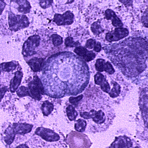
CSF. The large pale cell in the middle, among the white cells, is the amoeba - and finding one here is a medical emergency, not a routine report.
Source: CDC / DPDx | Public domain (U.S. federal government work) | Original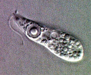
The flagellate form, which Naegleria switches into in water and only rarely shows in CSF. Acanthamoeba never does this at all.
Source: CDC / DPDx | Public domain (U.S. federal government work) | Original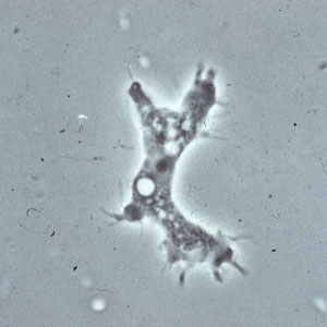
Acanthamoeba trophozoite. The fine spines radiating off it are acanthapodia, and they are what the genus is named for.
Source: CDC / DPDx | Public domain (U.S. federal government work) | Original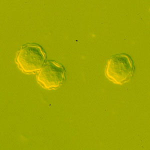
The cysts have two walls: a wrinkled outer one and a polygonal inner one. Naegleria cysts are smooth and single walled, and are not formed in human tissue.
Source: CDC / DPDx | Public domain (U.S. federal government work) | OriginalShown as published and free of charge from the source above, which does not endorse Learn Microbes. Full image credits
Source and morphology anchors
| Visual clue | Meaning | Student shorthand |
|---|---|---|
| Motile amoebae in CSF, recent warm freshwater exposure | Naegleria fowleri until proven otherwise | Primary amebic meningoencephalitis; notify immediately |
| Blunt lobopodia, fast directional movement | Naegleria pattern | Warm the slide slightly; cold slows the organism |
| Spine-like acanthopodia, sluggish movement | Acanthamoeba pattern | Think corneal scraping, contact lens case, or tissue |
| Double-walled cyst with polygonal inner wall | Acanthamoeba cyst | Naegleria does not form cysts in human tissue or CSF |
| Flagellate form after distilled water | Naegleria | Acanthamoeba and Balamuthia have no flagellate stage |
Free-living amoebae study readout
| Pattern | What to call it | How to think about it |
|---|---|---|
| CSF: motile trophozoites, lobopodia, no cysts | Presumptive Naegleria fowleri | Urgent call to the provider; follow the lab critical-value policy and reference testing |
| Cornea: spiny trophozoites or double-walled cysts | Acanthamoeba | Keratitis linked to contact lens wear and poor lens hygiene |
| Brain tissue: trophozoites and cysts, chronic course | Acanthamoeba or Balamuthia | Granulomatous amebic encephalitis; genus needs reference methods |
- Naegleria: CSF, lobopodia, flagellate form, no cysts in the patient.
- Acanthamoeba: cornea or tissue, spiny acanthopodia, double-walled cysts.
- Keep the CSF at room temperature, examine it quickly, and treat motile amoebae in CSF as an urgent finding.
Lobopodia in CSF = Naegleria. Spines and double-walled cysts = Acanthamoeba. Never refrigerate the specimen.
New bench cards and study tools are added regularly. Join the Lab List to hear about new cards and have a say in what gets built next.