
Chilomastix mesnili: Lemon-Shaped Cyst and Trophozoite
Original bench-card visuals for Chilomastix mesnili, a nonpathogenic intestinal flagellate: a small lemon-shaped cyst with an anterior knob, and a pear-shaped trophozoite with a spiral groove and a prominent cytostome.
Chilomastix mesnili: lemon-shaped cyst and pear-shaped trophozoite
Cyst: about 6-10 um, lemon or pear shaped with a clear anterior knob, one nucleus, and a curved cytostomal fibril often compared to a shepherd's crook. Trophozoite: about 6-24 um, pear shaped, one nucleus, a spiral groove along th...
About 6-10 um. The clear knob at one end gives the lemon shape. One nucleus. On a permanent stain the curved cytostomal fibril beside the nucleus looks like a shephe...
About 6-24 um. Pear shaped with one anterior nucleus. Three free flagella at the front and one inside the cytostome. A spiral groove gives the body a twisted look, a...
What this looks like down the scope
The board above is drawn to isolate the pattern. These are clinical images of the same stages, published as you see them, so you can find the pattern with everything else still in the field.
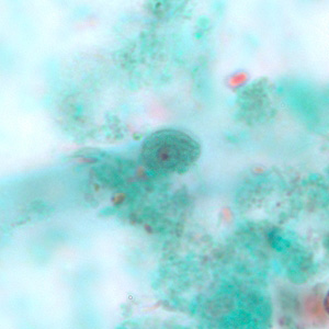
Lemon shaped with a small nipple at one end. That outline is the whole identification - it looks like nothing else in stool.
Source: CDC / DPDx | Public domain (U.S. federal government work) | Original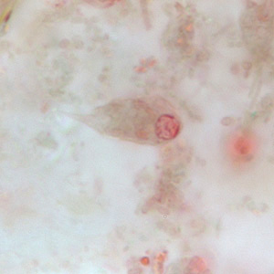
The trophozoite, pear shaped with a spiral groove, the cytostome, cut into one side. Non-pathogenic, but it means the patient swallowed something they should not have.
Source: CDC / DPDx | Public domain (U.S. federal government work) | Original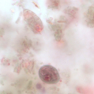
Both stages in one field, cyst below and trophozoite above, which is a useful size and shape comparison in a single frame.
Source: CDC / DPDx | Public domain (U.S. federal government work) | OriginalShown as published and free of charge from the source above, which does not endorse Learn Microbes. Full image credits
Chilomastix versus Giardia anchors
| Visual clue | Meaning | Student shorthand |
|---|---|---|
| Lemon-shaped cyst with a knob | Chilomastix mesnili cyst | Giardia cysts are oval with no knob |
| One nucleus in the cyst | Chilomastix mesnili | A mature Giardia cyst has 4 nuclei |
| Shepherd's crook fibril | Cytostome support in Chilomastix | Giardia shows axonemes and median bodies instead |
| Pear-shaped trophozoite, one nucleus, spiral groove | Chilomastix trophozoite | Giardia has two nuclei and a sucking disk |
| Stiff rotary motility | Chilomastix | Giardia is described as falling-leaf motility |
Chilomastix mesnili identification guide
| Pattern | What to call it | How to think about it |
|---|---|---|
| Lemon-shaped cyst, 6-10 um, one nucleus | Chilomastix mesnili cyst | Nonpathogen; report per SOP |
| Pear-shaped trophozoite, one nucleus, large cytostome | Chilomastix mesnili trophozoite | Confirm on permanent stain |
| Oval cyst, 4 nuclei, axonemes | Giardia duodenalis | Pathogen; see the Giardia bench card |
- The lemon-shaped cyst with an anterior knob is the classic picture.
- One nucleus and a large cytostome separate it from Giardia in both stages.
- It is a commensal, but it still tells you the patient was exposed to contaminated food or water.
Lemon-shaped cyst + one nucleus + shepherd's crook = Chilomastix mesnili, not Giardia.
New bench cards and study tools are added regularly. Join the Lab List to hear about new cards and have a say in what gets built next.