
Balantioides coli (Balantidium coli): Ciliated Trophozoite and Cyst
Original bench-card visuals for recognizing Balantioides coli, formerly Balantidium coli, in stool: a very large ciliated trophozoite with a kidney-shaped macronucleus, and a round thick-walled cyst.
Balantioides coli: ciliated trophozoite and thick-walled cyst
The largest protozoan found in human stool and the only ciliate that causes human disease. Trophozoite: about 40-200 um, oval, covered in short cilia, with a kidney-shaped macronucleus. Cyst: about 50-70 um, round, thick wall, mac...
About 40-200 um, oval, with rows of short cilia over the whole surface. A funnel-like cytostome sits at the narrower anterior end. The large kidney-shaped macronucle...
About 50-70 um, round to slightly oval, with a thick refractile wall. The macronucleus remains visible. Cilia may be seen inside young cysts but are lost in older on...
What this looks like down the scope
The board above is drawn to isolate the pattern. These are clinical images of the same stages, published as you see them, so you can find the pattern with everything else still in the field.
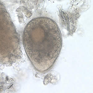
Enormous by protozoan standards, and covered in cilia. Nothing else in a stool prep is this big - that alone usually makes the call.
Source: CDC / DPDx | Public domain (U.S. federal government work) | Original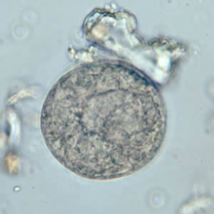
The cyst: round, thick walled, still very large. The bean-shaped macronucleus inside may or may not be visible depending on focus.
Source: CDC / DPDx | Public domain (U.S. federal government work) | Original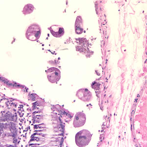
Colon tissue, H&E. This organism invades the bowel wall, so biopsy is a legitimate route to the diagnosis when stool is negative.
Source: CDC / DPDx | Public domain (U.S. federal government work) | OriginalShown as published and free of charge from the source above, which does not endorse Learn Microbes. Full image credits
What to look for in stool
| Visual clue | Meaning | Student shorthand |
|---|---|---|
| Very large oval organism on low power | Think Balantioides coli | No other intestinal protozoan reaches this size |
| Cilia covering the surface | Ciliate, not an amoeba or flagellate | The only ciliate that infects humans |
| Kidney-shaped macronucleus | B. coli nuclear clue | Seen in both the trophozoite and the cyst |
| Rotating, boring motility | Live trophozoite in fresh stool | Motility is lost quickly, so examine liquid stool promptly |
| Round thick-walled body with one large nucleus | B. coli cyst | Compare with helminth eggs of similar size before reporting |
Balantioides coli identification guide
| Pattern | What to call it | How to think about it |
|---|---|---|
| Large ciliated trophozoite with kidney-shaped macronucleus | Balantioides coli trophozoite | Report per lab SOP, giving the current and former name if your lab does so |
| Round thick-walled cyst, 50-70 um, macronucleus visible | Balantioides coli cyst | Measure with a calibrated ocular micrometer and compare with helminth eggs |
| Dark, featureless oval body on trichrome only | Not enough to report | Go back to the wet mount or concentrate to confirm cilia and nucleus |
- Largest intestinal protozoan and the only ciliate pathogenic to humans.
- Cilia plus a kidney-shaped macronucleus identify it; the micronucleus is rarely seen.
- Use wet mounts and concentrates. Permanent stains are too dark to be reliable for this organism.
Huge, ciliated, kidney-shaped macronucleus = Balantioides coli. Recognize it on the wet mount.
New bench cards and study tools are added regularly. Join the Lab List to hear about new cards and have a say in what gets built next.